USES:
The model is designed as a visual aid for teaching physiology and hygiene courses.
It helps the students to understand the anatomical structure of the head, neck and
Internal organs of the human body.
DEMONSTRATIONS:
The model shows the relative positions, morphological characters, and anatomical
Structures of the head, neck, and internal organs with special reference to respiratory,
Digestive, urinary, and nerve systems.
The head and neck
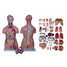
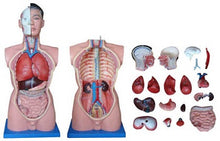
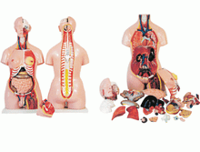
The right side of the head shows partially the skull bones and chewing muscles (Masseter and temporal muscles). The 12 pairs of cranial nerves are distinctly shown On the ventral side of the brain. The eyeball irremovable. The head and neck can Be used for demonstration of the nasal cavity and mouth cavity. The laryngeal part Shows the cavity of the larynx, sinus of the larynx, and glottal Rima. The parathyroid gland Can be seen at the posterior board of the lateral lobe of the thyroid gland.
The thorax and abdomen
The right and left lungs are divided into two lobes each to show the structure of The hilum. A coronary section of the heart displays the structural differences between the right and left atria, and between the right and left ventricles. Large blood vessels such as the superior and inferior vena Cara, pulmonary arteries, and aorta are shown also. Internal organs including the liver, stomach, pancreas, intestines, spleen, kidney, urinary bladder, etc. are situated in the abdominal cavity underneath the diaphragm. The descending part of the duodenum, caecum, and a small portion of ileum and jejunum show the structure of the wall of the alimentary tract. The dissecting surface of the right kidney reveals the structures of the renal cortex, renal medulla, and renal pelvis. The model also features an exposed spine with removable vertebra and spinal cord the segment, a female breastplate, and interchangeable male and female genitalia.
CONSTRUCTION: The model is made of PVC plastic. On base.
SIZE: 85CM tall.


