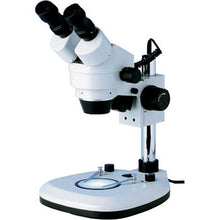
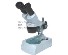
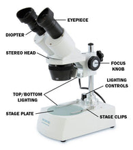
Applications:
Widely used in the electronic industry, assembling, and inspection of precision instrument and meter, educational experiment, observation, and research. It can be used in schools, research institutes, factories, and families to study geology, the outer appearance of objects.
Major technical index:
Optical index(mm)
Field of view
Working distance
Electrical Index :
Input voltage:220V/50Hz or l 10V/60Hz(Optional)
- illuminating style:
- natural light
- incident light 12V/10W(without transmitted illuminator)
- halogen light 12V/10W(without transmitted illuminator)
- incident and transmitted illuminator 12V/10W
- incident and transmitted halogen illuminator 12V/10W
structure Index :
Vertical binocular bead, Slanting 45° binocular head is optional. Diopter adjustment +/-5°, inter-pupillary distance adjustable 54-76mm.
How to use:
1) environment requirement: Dry dust-free room temperature between -5- +40 degrees Celsius.
2) Illuminator control:
Plugin the power cord into the outlet. Refer to the following table for illumination styles. For microscopes with dimmer control, the brightness of the illuminators can be adjusted.
3) Select the stage:
1) The frosted glass stage is placed on the base and is fixed with a screw, it is used when a transparent specimen is being observed, please use the transmitted illuminator.
2) Black and white stage is kept in the packing as an accessory. When it is used, please take off the glass stage and place the black and white stage on the base. Normally the white side is upward. If the specimen is White or in other bright colors, use the black side to improve the contrast with the only incident illuminator.
4) Placement of specimen:
Place the clean specimen on the center of the stage and fix it with clipper if necessary.
5) Use of rubber eye-guard: One pair of rubber eye guards is contained in the packing. They are used to protect against incident light around the eyepieces to improve visibility.
6) Focusing, diopter, inter-pupillary adjustments:
Place a specimen onto the stage. Loosen the body locking thumbscrew and bold the microscope head and move the body up and down and fix it at the estimated working distance. Rotate the zooming knob while looking through the right eyepiece until you see the image. Using the focusing handles to get the sharpest image of the specimen. Then look through the left eyepiece with your left eye and turn the diopter adjustment ring until you get an image as sharp as the right side. Make this adjustment without moving the focusing handle. Then grasp the right and left prism housing and move them closer or further apart in order to match your pupil distance. Adjustment is proper when the field of view becomes comfortable and presents a full single field.
7. Objective power switch :
Models of XTX-3 have 180 ° switch objectives.
5. Replacement of bulbs and fuse:
Warning: Always disconnect the power cord when you change the lamp or fuse and make sure that the lamp is not hot.
1- Replacement of the incident lamp:
Loosen the fixing screw of the shade and take off lamp hosing. Replace the bulb with the same new bulb. Place the lamp housing back and fix it with the same screw.
2- Replacement of the transmitted lamp:
Loosen the fixing screw of the glass stage and take off the glass. Takedown the broken bulb through the stage bole and install a new bulb.
3- Replacement of the fuse:
The fuse case is located at the backside of the base. Unscrew the fuse case cover and put in a new one.
6, Maintenance and general care of your microscope
- A microscope is a delicate precision instrument and it may be damaged by dropping and hitting.
- Do not keep a microscope under the sun. It should be kept in a dry and clean environment and avoid beat and strong tremor.
- To obtain a clear image, do not touch lenses with your finger.
- All lens surfaces should be kept clean. Lf the lens get dusty blow off the dust with a rubber syringe. If necessary clean the lenses with lint-free cloth dipped in the aether.
- Do not use any organic material to clean the microscope surface, especially the plastic surface. It should be cleaned by neutral detergent.
- Because the assembly of all parts has been done by skilled optical craftsmen at the factory, you should never attempt disassembly.
- Apply a little bit grease regularly to the mechanical parts. . .
- When not in use always cover the microscope with the dust cover and place it in a cool and dry place.
Optional parts:
- eyepieces: (table omitted)
- Darkfield stage, Jewel twee1.Cf'S, particularly for jewel inspections.
- Ring lamp: The fluorescent lamp is fixed by 3 screws on the outside surface of the objective case. It can be a substitute for the traditional incident lamps because it is evenly hogtied and the light is brighter and softer and more comfortable to use.


